
About Dr. D. (Daan) Brinks, PhD

Daan Brinks studied Applied Physics at Twente University. He obtained his PhD in Molecular Nanophotonics with Niek van Hulst at ICFO – The Institute of Photonic Sciences in Castelldefels (Barcelona), Spain, working on the development of nonlinear microscopy methods to obtain detailed information from single biomolecules in physiologically relevant environments. He then obtained a Rubicon Grant from NWO to move to the department of Chemistry and Chemical Biology at Harvard University for a postdoc with Professor Adam Cohen. He combined work in optical and genetic engineering to resolve neural dynamics with high spatial and temporal resolution, for instance obtaining exceptionally rich data on electrical signal propagation in dendritic trees.
He worked as an HHMI postdoctoral fellow in the Cohen lab before joining Delft University in Technology in 2017 as an assistant professor in Imaging physics. Here he works on the development and application of cellular imaging and signal detection in neurobiology, cancer biology and developmental biology. In 2018, he also joined the department of Molecular Genetics at Erasmus MC. He has been awarded an NWO START-UP grant.
Visit Daan's lab
Introduction
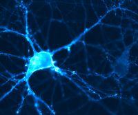
We address questions of cell function through optical perturbation ad imaging. We develop tools with roots in physics, biochemistry, optics, mathematics and nanofabrication and we're interested particularly in neural function work on every level, from biophysical principles to consequences in behavior and from subcellular compartments to complete organisms.
Research topics
We address questions in cell biology from an imaging perspective: How to transduce information from cells into detectable photons, resolvable in space and time.
We have a particular soft spot for nonlinear microscopy and Genetically Encoded Voltage Indicators (GEVIs), but happily use principles, tools and techniques from Physics, Chemistry, Mathematics, Molecular Biology, Nanofabrication and Photonics where applicable.
Current projects include:
Voltage Nanoscopy
Signal processing in neurons begins and ends at synapses. Voltage imaging, with its promise of resolving membrane voltage dynamics in space and time, is the ideal candidate to resolve fast voltage dynamics at synapses and neuromuscular junctions, but is limited by the optical diffraction limit and finite photon fluxes.
We optically investigate voltage dynamics at dendritic spines and axon terminals using plasmonically enhanced voltage imaging. The excitation and emission of molecules can be enhanced by electromagnetically coupling the molecule to a plasmonic particle, as an optical antenna. An added advantage is that this enhancement takes place in the near field of the optical antenna, leading to nanoscopic areas of excitation and emission enhancement and pushing voltage imaging beyond the boundaries of the optical diffraction limit. We apply this method to suitable voltage sensing proteins or dyes, embedded in the cell membrane at synapses or neuromuscular junctions. We are interested in whether plasticity can manifest as varying voltage dynamics in these subcellular structures.
Absolute Voltage Imaging
In a proof of principle paper we introduced 2-Photon Absolute Contrast Lifetime Imaging Voltage Sensing (2PACLIVS) as a method to optically obtain absolute membrane voltage information. This method relies on Genetically Encoded Voltage Indicators (GEVIs), engineered such that membrane voltage influences the fluorescence lifetime of the probe. In this project we aim to create an optimized probe and optical setup for absolute voltage imaging, and apply the technology to to look at embryonic development of Zebrafish.
Multiphoton Voltage Imaging
We explore the possibilities of genetic engineering and optimization of optical microscopy to achieve robust, deep tissue voltage imaging using multiphoton excitation. We're interested in how to record optical signals from a large number of cells, all at different 3D coordinates, in scattering tissue, in a moving background, in the limited window available to sustain the time resolution to resolve action potentials.
