
About our research group/lab
Our research
What is the motivation for this research?
A heart attack occurs when the flow of blood in the coronary artery is blocked. This is mostly due to the development and rupture of vulnerable atherosclerosis plaque. Is there a way to capture those vulnerable plaques at early stage and thus see the heart attack before it happens?
What is the aim?
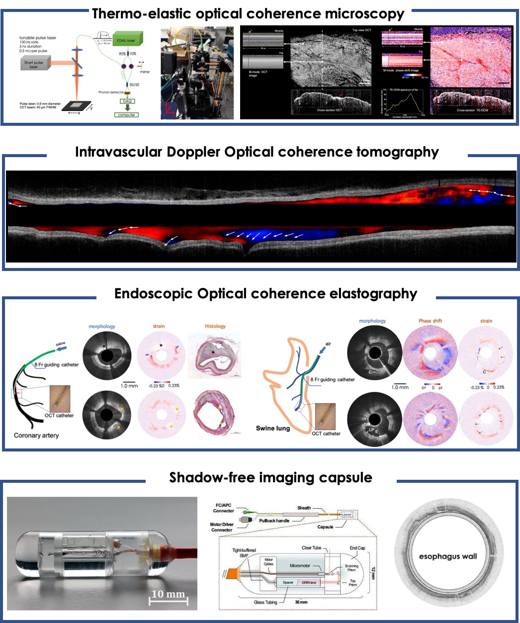
In this research, we will develop three new imaging modalities to visualize four different types of properties of the coronary artery, i.e. microstructure, flow pattern, thermo-elastic property and stiffness. By doing so, comprehensive accessment of morphology and physiology of the coronary artery are provided, which may bring new opportuties for the early detection of vulnerable plaques. The new technologies developed in this project may also be used for the ealy diagnosis of other dieases in the future.
How will you perform this research?
- Thermo-elastic Optical Coherence Tomography: By detecting the tiny tissue displacement induced by a nanosecond laser pulse, we can create a new modality, showing both the thermo-elastic properties and microstrcutures of tissue.
- Megahertz intravascular Doppler OCT: Advancing the Doppler OCT technique to 1.5 Megahertz speed allows us to measure flow velocity at <37.5 cm/s, and further visualize flow pattern of entire coronary artery.
- Endoscopic Optical Coherence Elastography (OCE): Using a stable catheter and a fast OCE system, we will make the first endoscopic realizationg of OCE, simultaneously mapping the stiffness and structure of coronary artery (and bronchus).
What is the desirable outcome?
Technical outcome:
- Microscopic TE-OCT device for pathological diagnose
- Handheld TE-OCT for skin imaging.
- Clinical compatible intravascular Doppler OCT device.
- Endoscopic stiffness imaging for coronary artery, bronchus and esophagus.
Biomedical outcome:
- Visualizing the lipid signature of vulnerable plaque using TE-OCT
- Mapping the mechanical properties of plaque and predicting the vulnerability
- Visualizing the flow pattern of whole artery and offering new information to PCI procedure
Collaborations
Collaborations within Erasmus MC
- Department of cardiology
- CoFa EMI
- Department of respiratory medicine
- Department of gastroenterology and hepatology
- Department of pathology
Collaborations outside Erasmus MC
- Universität zu Lübeck, Biomedizinsche Optik, Huber lab
Funding & Grants
NWO Veni 2017 - 15940