
What we do
About our project
Motivation for this project
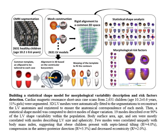
Cardiovascular disease (CVD) has been the leading cause of death in almost every region of the world for decades, despite better detection and treatment. This mandates a shift towards personalised and preventive CVD management. 3D geometric modelling is a new method of analysing cardiac magnetic resonance data, thereby enabling to model and compare the cardiac ‘shape’ of individuals. Applying this new method on paediatric cohorts could help understand the ‘healthy’ shape of a child’s heart and its response to risk factor exposures. This knowledge may have implications for screening, prevention and interpretation of the influences of CVD risk factors.
Explanation of the project
This project applies 3D geometric modelling to assess how children’s cardiac shape influenced by early life CVD risk factors such as childhood obesity, preterm birth and maternal obesity. The accuracy and predictive power to detect subtle influences on cardiac shape depend on the size and quality of a control group. We will first determine the range of normal variation in cardiac geometry in 2631 healthy children (age 10.2±0.6 years). This will generate reference values against which to compare values of subgroups with CVD risk factors. For example, the heart of obese children showed septo-lateral tilting, antero-posterior compression, and decreased eccentricity.
Potential impact of the project
This project will add to the Cardiac Atlas Project to establish a shareable, web-accessible, structural and functional atlas of the normal paediatric heart. This will facilitate collaborative statistical analysis of the paediatric heart shape and comparison of cardiac shape and function among and within paediatric groups. Second, the project includes assessing the effects of a maternal preconception lifestyle intervention on the offspring’s cardiac shape. If it appears that the intervention has the desired effect, future CVD policies to reduce cardiovascular morbidity and mortality in generations to come may focus on optimizing maternal lifestyle before and during pregnancy.
Our research focus
Early life risk factors of CVD
Epidemiological studies have shown a relationship between foetal or neonatal insults, and the development of adult diseases such as CVD, hypertension, type 2 diabetes and obesity Maternal obesity increased the risk of obesity and metabolic dysregulation in the offspring more than three-fold. Preterm birth carried a 30% higher risk of cardiovascular mortality in a cohort of 674,820 young adults, and a 17-fold higher risk of heart failure in adolescents. Because in Western countries 60% of women of reproductive age are overweight or obese. and 10% of births are preterm, any related adverse health impact is relevant to a large population.
Geometric Analyses
Cardiac Magnetic Resonance (CMR) allows accurate, non-invasive assessment of the structure and function of the adult and the paediatric heart. Traditional outcome measures such as ventricular mass, cardiac dimensions or cardiac output fail, however, to capture the wealth of data generated by CMR examinations. The technique of ‘3D geometric modelling’ can capture variations of 3D cardiac shape within populations, and can provide detailed information on the mechanisms of disease processes to explore the early manifestations of CVD risk factor influences in early life.
This project
This project will raise the potential for improving maternal and perinatal care by identifying biomarkers for the surveillance of individuals exposed to early-life risk factors, and for new early prevention strategies to complement classical CVD management. Second, this proof-of-principle study is the first to apply geometric CMR analyses in children to identify changes in cardiac geometry that may reflect detrimental cardiovascular health. Later it could be applied in other paediatric scenarios, e.g. congenital heart disease. Third, we will develop a shareable, web-accessible, atlas of normal geometry of the paediatric heart, based on data of a large cohort of healthy children.
Funds & Grants
This project is funded by:
- ZonMw Rubicon grant 452183001;
- Netherlands Heart Institute Fellowship grant;
- European Society of Cardiology Research grant.
Collaborations
Collaboration within Erasmus MC
- Department of Paediatrics, Subdivision of Paediatric Cardiology, Sophia Children’s Hospital;
- Generation R Study Group;
- Department of Epidemiology.
Collaboration outside Erasmus MC
- Oxford Cardiovascular Clinical Research Facility, Division of Cardiovascular Medicine, Radcliffe Department of Medicine, University of Oxford, Oxford, United Kingdom;
- Department of Biomedical Engineering, School of Biomedical Engineering and Imaging Sciences, Kings’ College London, London, United Kingdom.
Our team
Principal Investigator:
- Arend W. van Deutekom, MD PhD, paediatric cardiologist.
e-mail
Team members:
- Prof. W.A. Helbing, MD PhD, professor in paediatric cardiology. Head of paediatric cardiology;
- Prof. V.W.V. Jaddoe, MD PhD, Professor of Pediatrics- Epidemiology. Head of Generation R study group.
