
About our research group/lab
Background
As many drugs have difficulty reaching their site of action to effectively treat diseases, drugs are given in high dosages causing serious side-effects. Toxic effects to the rest of the body can be prevented by local drug delivery. Ultrasound-activated microbubbles, 1-10 µm in diameter, have shown potential as local drug delivery system. When exposed to ultrasound, the microbubble’s gas core responds to the pressure change by compressing and expanding, which results in the vibration of the microbubble. This vibration stimulates and enhances cellular drug uptake. The group studies the underlying mechanism of the microbubble-cell-drug interaction to achieve maximum therapeutic outcome.
Overall aim
As with all local drug delivery systems, the aim is to achieve a specific pharmacological response of a therapeutic agent at a particular diseased site. The benefits are a more controlled biodistribution of the therapeutic agent which reduces side-effects and improves therapeutic efficacy. A unique feature of ultrasound is local insonification thereby stimulating and enhancing drug uptake only at a region of interest. In addition, ultrasound imaging will aid the guidance and monitoring of therapy. Given the presence of microvascular networks in nearly all tissues, local drug delivery using microbubbles provides extensive possibilities for treating pathological tissues.
Research focus area
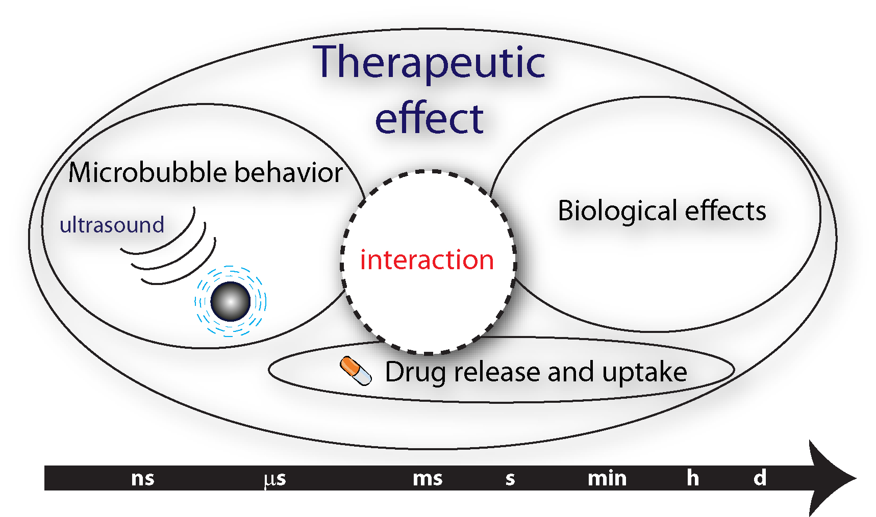
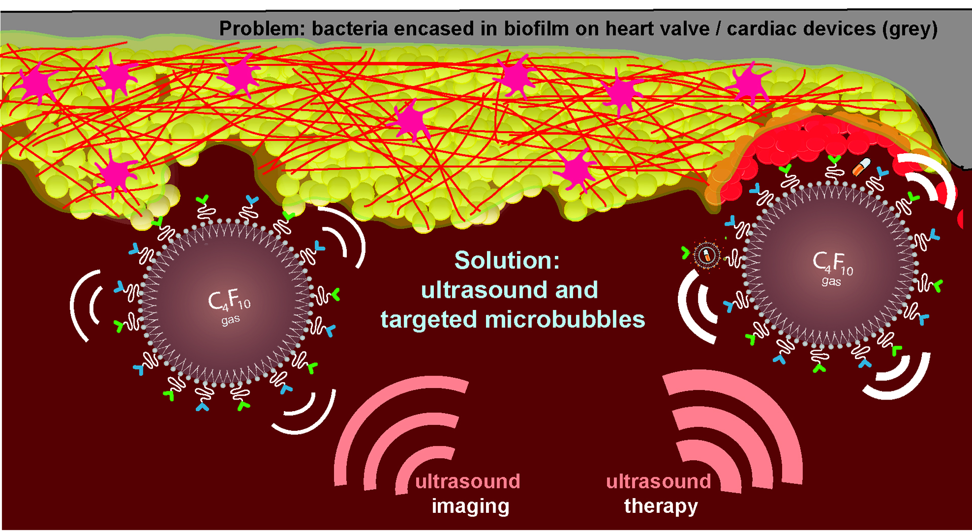
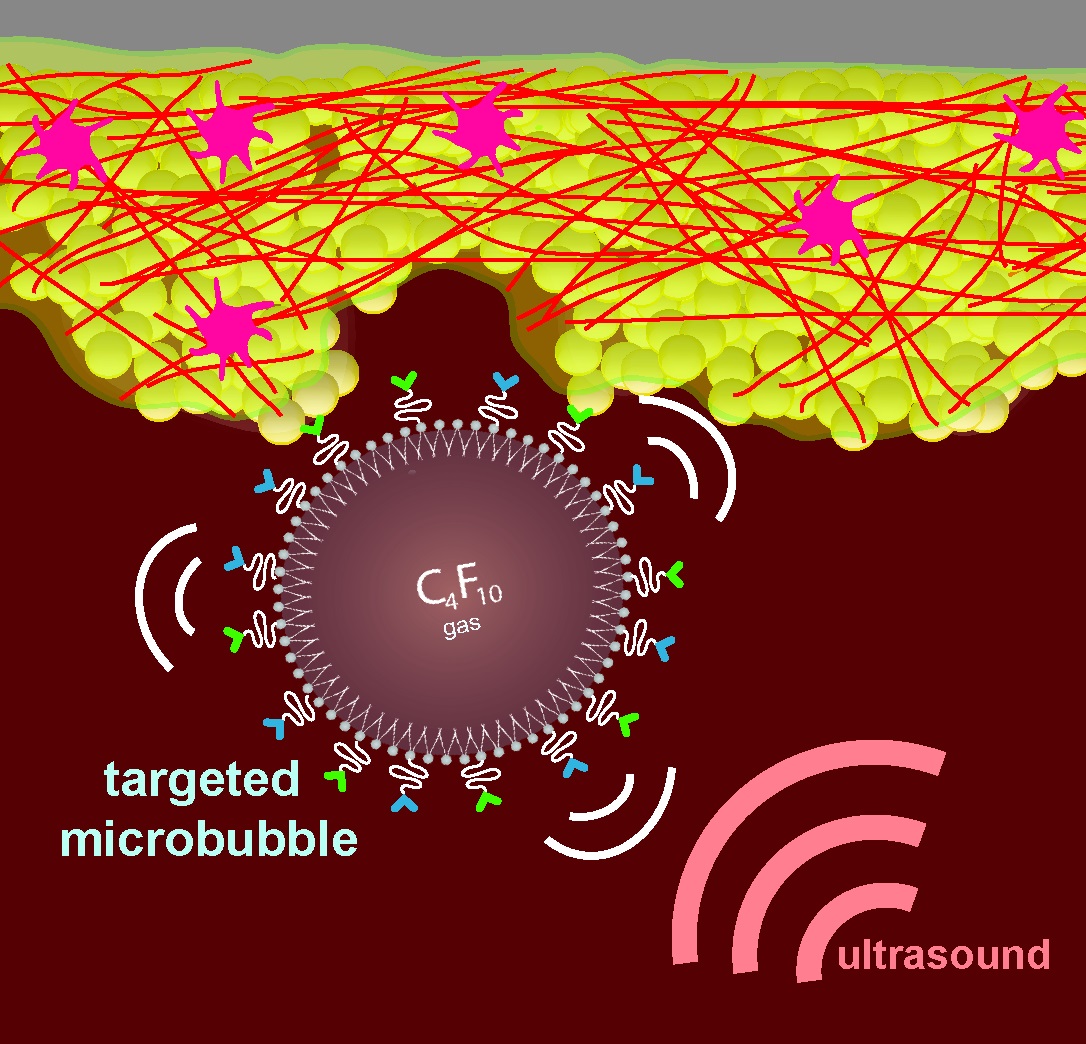
We focus on producing novel coatings of microbubbles for imaging and drug delivery, elucidating the microbubble-vessel wall-drug interaction, and developing microbubble-mediated treatment and ultrasound molecular imaging of bacterial infections on heart valves/cardiac devices. Detailed insight into the underlying mechanism of the microbubble-cell-drug interaction is crucial for maximum therapeutic outcome and widespread clinical use. The different time scales involved makes this research challenging, namely nanoseconds for the the microbubble vibration (microbubbles vibrate 2 million times per second in a 2 MHz ultrasound field), milliseconds for physiological effects, seconds to minutes for biological effects, and days to months for clinical relevance.
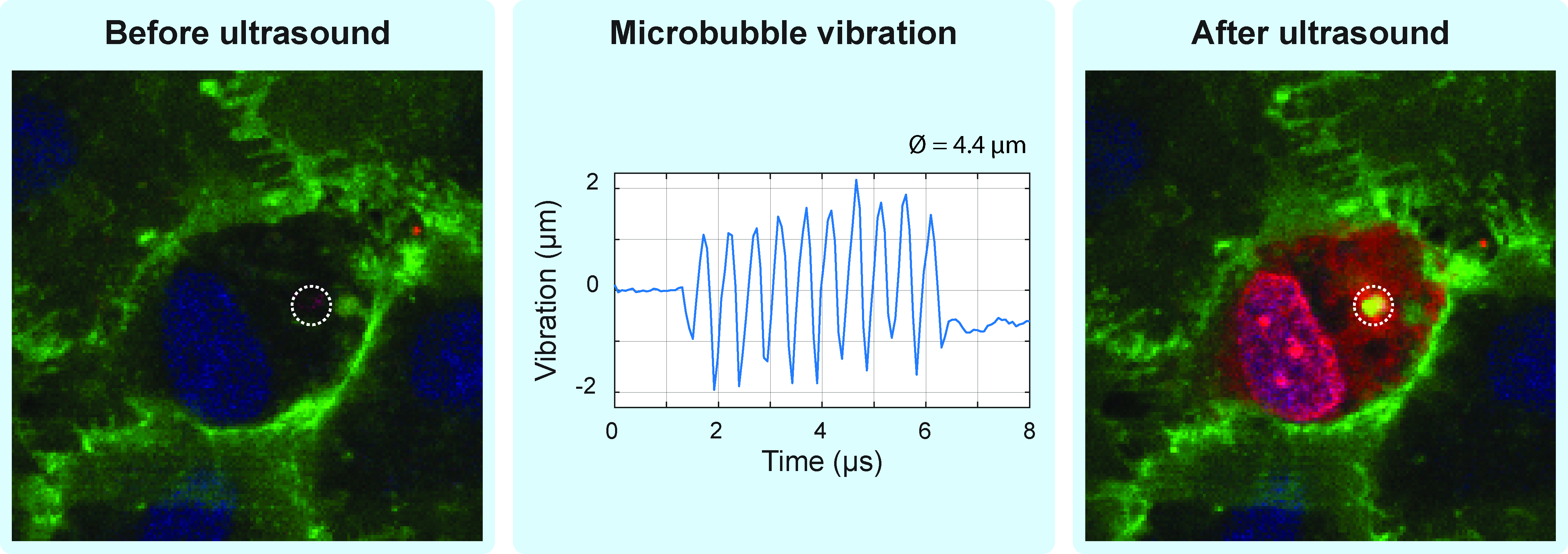
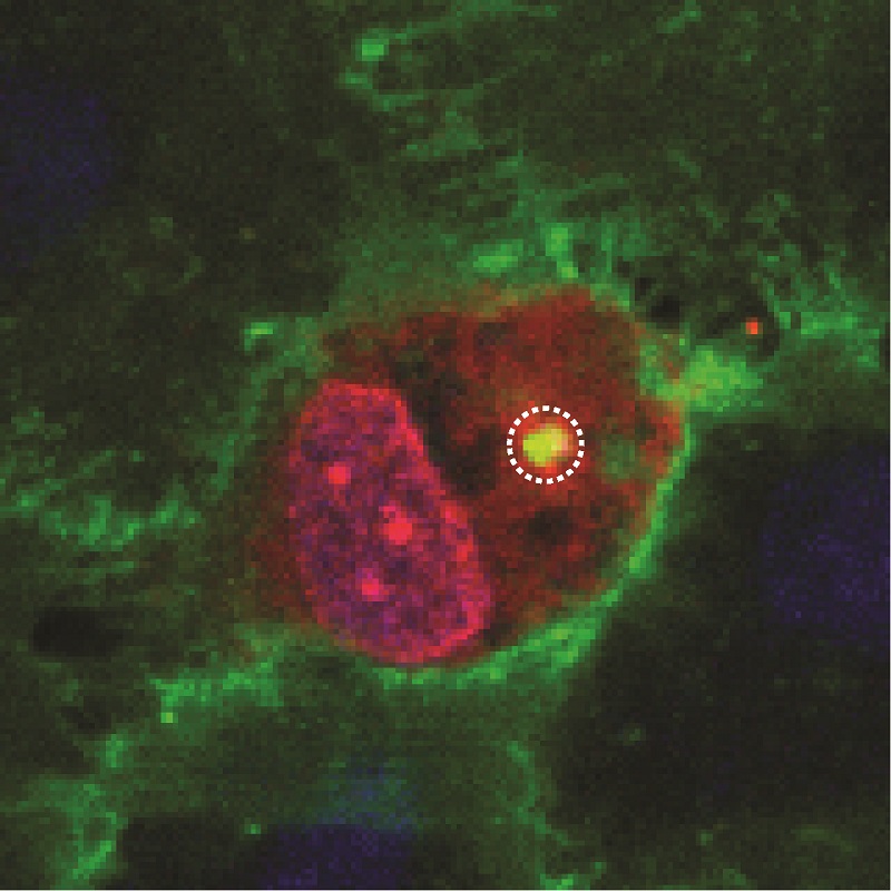
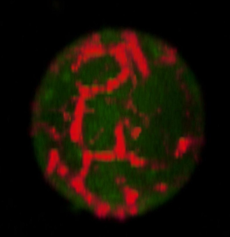
Confocal microscopy images of endothelial cells before and after the vibration of a microbubble by 2-MHz ultrasound. After ultrasound, the model drug (red) is taken up by the endothelial cell. The microbubble location is indicated by the dotted line and the cells are stained in green (cell membrane) and blue (cell nuclei).
Coupling Two Ultra-high-Speed Cameras to Elucidate Ultrasound Contrast-Mediated Imaging and Therapy
Internalization of targeted microbubbles by endothelial cells and drug delivery by pores and tunnels
Dispersing and Sonoperating Biofilm-Associated Bacteria with Sonobactericide
Vancomycin-decorated microbubbles as a theranostic agent for Staphylococcus aureus biofilms
The Impact of Lipid Handling and Phase Distribution on the Acoustic Behavior of Microbubbles
Ultrasound-responsive cavitation nuclei for therapy and drug delivery
An in vitro proof-of-principle study of sonobactericide
Combined Confocal Microscope and Brandaris 128 Ultra-High-Speed Camera
Acoustic behaviour of microbubbles and implications for drug delivery
Collaborations within Erasmus MC
Cardiothoracic Surgery
Hematology
Medical Microbiology & Infectious Diseases (MMID)
Pathology
Collaborations outside Erasmus MC
Image-guided Ultrasound Therapeutics Laboratories, University of Cincinnati, Holland lab
University of Virginia, Klibanov lab
Physical Chemistry, Martin Luther University Halle-Wittenberg, Blume lab
Leeds Microbubble Consortium
Laboratory of Acoustical Wavefield Imaging, Delft University of Technology
- NWO Vidi 17543
- ERC Starting grant 805308
- Phospholipid Research Center, Heidelberg, Germany
- Erasmus MC fellowship
Klazina Kooiman , PhD, Principal Investigator
Assistant Professor:
- Kirby R. Lattwein, PhD
Technician:
- Anne J.Poos, BSc
Postdoctoral fellow:
- Gonzalo Collado-Lara, PhD
PhD students:
- Bram Meijlink, MSc
- Hongchen Li, MSc
- Yuchen Wang, MSc
Alumni:
- Mariël Leon – Grooters, BSc (Aug 2019 - July 2024)
- Joop J.P. Kouijzer, PhD obtained on 15 November 2023
- Simone A.G. Langeveld, PhD obtained on 10 May 2022
- Inés Beekers, PhD cum laude (with distinction) obtained on 2020-07-08; PhD student 2015 – 2019; postdoctoral fellow 2019-12 – 2020-11.
- Reza Pakdaman Zangabad, PhD, postdoctoral fellow 2020-03 – 2021-12