
Services
Erasmus MC has been investing in imaging expertise and systems for quite some time and has built unique expertise in a number of areas. Various technologies (i.e. equipment as well as expertise) are put together in one new facility: Applied Molecular Imaging Erasmus MC. Combinations of imaging modalities can be applied to a single research question, or one experimental animal. From experience we know that a physical grouping of complementary techniques and expertise is a must to reach successful synergy. The new central imaging facility is largely housed in the new research unit in the Erasmus MC Experimental Animal Centre (EDC).
The research in the departments of Erasmus MC covers a wide range of topics; cancer research, microbiology/immunology, developmental biology and pharmacology.
Discover our comprehensive selection of state-of-the-art preclinical imaging equipment designed to enhance your research capabilities:
- Discovery MR901 – 7T MRI
- FMT2500 – Fluorescent Optical Imaging
- Gammacounter for radioactive samples
- IVIS Spectrum – Optical Imaging
- MSOT – 3D Optoacoustics
- NanoScan – SPECT, 1T MRI
- Quantum Gx2 – microCT
- VECTor – SPECT/PET, microCT and Optical Imaging
- Vevo3100 - Ultrasound
- Vevo LAZR-X photoacoustic addition to the Vevo3100
- Workstation with analysis software packages Imalytics, PMOD, Analyze, VEVOlab and more
Explore our showcases of cutting-edge preclinical imaging, where groundbreaking techniques and applications come to life, advancing our understanding of complex biological processes and disease mechanisms.
1. Quintuple Modality Tumor Necrosis Imaging
"Unlocking Insights into Tumor Aggressiveness and Therapy Effectiveness"
Leverage the power of both nuclear and optical imaging to pinpoint therapy-induced tumor necrosis. This multimodal approach offers complementary insights into tumor response. PET with FDG closely monitors tumor metabolism and serves as a benchmark for treatment efficacy. While SPECT and fluorescent imaging (FLI), armed with an advanced necrosis-avid contrast agent, accurately reveal areas of necrosis, exploiting damaged membrane integrity to image cell death. Additionally, bioluminescent imaging (BLI) detects viable tumor cells. Together, this comprehensive array of imaging modalities provide vital insights guiding tailored treatment strategies.

This figure was originally published in:
Stroet MCM, de Blois E, de Jong M, Seimbille Y, Mezzanotte L, Löwik CWGM, Panth KM. Improved Multimodal Tumor Necrosis Imaging with IRDye800CW-DOTA Conjugated to an Albumin-Binding Domain. Cancers. 2022; 14(4):861.
2. Optical Imaging of Tumor Response
“Explore the Impact of Therapy on Tumor Vasculature”
Optical imaging provides a non-invasive way to monitor processes involved in tumor growth and metastasis formation in immunodeficient mice. Here fluorescence molecular tomography (FMT), with its advanced 3D imaging capabilities, is used to delve into the effects of hyperbaric oxygen therapy (HBOT) and irradiation on tumor dynamics. By analyzing blood vessel quality with AngioSense and unraveling hypoxia with HypoxiSense probes, FMT offers a comprehensive overview of the tumor environment. Seamlessly integrating FMT with CT images provides precise anatomical references. Thus, optical imaging methods can unveil longitudinal insights into tumor characteristics, revolutionizing oncology research.
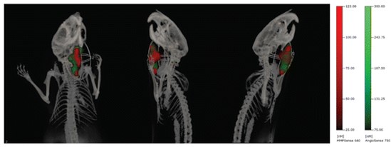
Braks JAM, Spiegelberg L, Koljenovic S et al. Optical Imaging of Tumor Response to Hyperbaric Oxygen Treatment and Irradiation in an Orthotopic Mouse Model of Head and Neck Squamous Cell Carcinoma. Mol Imaging Biol 17, 633–642 (2015).
3. Imaging Drug Delivery with SPECT and DCE-MRI
“Unveiling Tumor Dynamics”
The combination of SPECT and MRI can unveil vital insights into the perfusion efficiency and the accumulation of radiolabeled receptor-targeting peptides in solid tumors. This multimodal approach reveals nuanced variations in functional tumor characteristics, even in seemingly homogeneous tissue. By leveraging DCE-MRI-derived parameters, it is possible to decode tumor perfusion, vessel density, and permeability, unlocking insights into the impact of vascular traits on the efficacy of receptor-targeting therapies. 
This figure was originally published in:
Haeck JC, Bol K, de Ridder CMA et al. Imaging heterogeneity of peptide delivery and binding in solid tumors using SPECT imaging and MRI. EJNMMI Research. 2016; 6(3).
Access to and use of the AMIE instrumentation is permitted under the supervision of AMIE personnel. Training is provided for those who plan to use the equipment independently.
Costs for equipment use depends on user requirements. For more information contact the AMIE.
The AMIE provides full-service support on all equipment for imaging experiments, with the option to obtain training for independent use.
All imaging modalities have isoflurane equipment for anesthesia.