About Dr.ir. J.G. (Hans) Bosch, PhD
Introduction
Dr.ir. J.G. (Hans) Bosch is Associate Professor (fulltime, since 2011), staff member and Principal Investigator in the Thoraxcenter Biomedical Engineering group. He specializes in innovative 2D and 3D ultrasound image generation, processing/analysis and transducer development. Main research interests are new high-framerate imaging techniques and their application possibilities for blood flow, tissue motion, stiffness and function, as well as design and development of new transducers, imaging schemes and analysis methods.
Current Research:
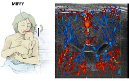
Ultrasound imaging is entering a new phase where advancing technology opens completely new paths. We are going from 2D imaging to fully 3D in many different applications, thanks to new, highly complex transducers with integrated electronics and thousands of active elements. Novel plane-wave imaging schemes allow a hundred-fold increase in the achievable imaging rates, allowing us to see very fast phenomena such as shear waves in the heart, the motion patterns of contrast bubbles in the blood flow, or the electrical activation of the heart wall. Much more sensitive detection of blood flow (e.g. in the neonatal brain) or tissue stiffness (heart wall in heart failure, diseased vessel walls) is becoming possible. We can even visualize the response of brain areas (functional ultrasound) to a stimulus.
Field(s) of expertise
- Advanced 2D and 3D ultrasound image generation
- 2D/3D image processing and analysis
- Design and development of advanced transducers, imaging schemes and analysis methods.
- High-framerate ultrasound imaging for blood flow, tissue motion, stiffness and function.
Education and career
M.Sc. degree in Electrical Engineering, Eindhoven University of Technology, 1985:
‘Speech recognition for application in aids for the disabled’
Ph.D. degree Leiden University Medical Center, 2006:
‘Automated analysis of echocardiographic images’
Promotors: Prof. J.H.C. Reiber, Prof. M. Sonka (Univ. Iowa)
Coming from a background in medical electronics (TU/e), he has first specialized in automated ultrasound image analysis, as a section leader Echocardiography and Assistant Professor at the Division of Image Processing (LKEB), Department of Radiology, Leiden University Medical Center, up to 2005. This has resulted in several practical methods for echocardiographic image analysis. In his current position, he has mostly specialized in new imaging techniques, image analysis and transducer development for 3D ultrasound and high-framerate imaging. All these projects aim at realizing clinically applicable technology and methods that provide new, unseen functionality. He closely collaborates within Medical Delta with colleagues at TU Delft in about 6 shared projects, and on a national level with most PIs within the Dutch ultrasound technology community, as well as on an international level.
Publications
Teaching activities
- Associate Professor, Thorax Biomedical Engineering, Cardiology
- Directly supervised PhD theses: 9 completed, 5 in progress
- Co-supervision of 6 more PhDs
- Opposition at ~25 (inter)national PhD defences
- Postdoctoral/PhD course organization and lecturing
- Seminar organization
- MSc/BSc supervision
Other positions
- Staff member, Thorax Biomedical Engineering group (BME), Department of Cardiology
- Head of section Advanced Ultrasound Imaging, BME (together with Rik Vos)
- Technical Committee, SPIE Medical Imaging, Conf Ultrasonic Imaging, 2010-now
- Organizer MICCAI CETUS challenge on 3D echocardiographic segmentation, Boston, 2014
- Conference Chair, SPIE Medical Imaging, Conf. Ultrasonic imaging, 2011-2015 (incl technical committee, session chairing etc)
Scholarships, grants, and awards
Grants:
Secured a large number (~20) of national and international grants as PI or co-PI, including STW/ NWO-TTW OTP, Hartstichting, Perspectief, NWO-Groot, EU.
Awards:
- Co- or last author of many award winning papers and conference proceedings
- Supervisor of two Cum Laude PhDs
- Many paper awards/fellowships/young investigator awards acquired by my PhD students

